

- Our doctors speak out
Patellar syndrome
Patellofemoral pain syndrome is defined as mechanical pain felt on the front of the knee. Compression activities such as squatting, climbing and descending stairs, or prolonged sitting can trigger or aggravate the syndrome. Repetitive activities such as running can exacerbate the pain to the point where it becomes impossible to continue. The pain subsides with rest and is reversible, although it can persist for quite some time after exertion, similar to the pain associated with iliotibial band syndrome, with which patellofemoral pain syndrome can be linked.
Patellofemoral pain syndrome primarily affects a young, active, or athletic population, more often female than male. In a specialized knee or sports medicine clinic, patellofemoral pain syndrome alone accounts for 25 to 40% of consultations.
Patellofemoral pain syndrome remains a subject of controversy to this day, with no valid explanation for its etiology. Predisposing factors such as constitutional hyperlaxity or significant femoral anteversion have been implicated, but it is a multifactorial problem. A functional disorder underlies the syndrome, and the explanation can only be found through movement analysis.
The loss of physiological inter-articular synchrony during gait is caused by a poorly understood and rarely investigated clinical entity: Functional Hallux Limitus (FHL). By systematically investigating for this condition over the past 20 years, I have observed that this entity is present and can be diagnosed in all cases of patellofemoral pain syndrome.
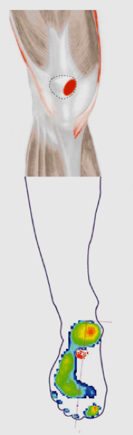
Pain may be the only symptom or be associated with sensations of giving way or catching, without necessarily indicating patellar instability with a fear of dislocation. Effusion is rare. The diagnosis is often made by exclusion, and radiological examinations generally show nothing abnormal, except perhaps transient edema of the patellar cartilage on MRI. No correlation has been established between cartilage lesions and pain, and the term "chondropathy patellae" is no longer used to define this syndrome, which does not progress to osteoarthritis.
From a therapeutic standpoint, there is no real consensus. Numerous studies have been published showing encouraging results with a comprehensive approach based on stretching and core strengthening. Foot supports sometimes have a beneficial effect. Unfortunately, the improvement is often temporary, and relapses are frequent. Without regular training, the pain returns, forcing patients to change their exercise habits and lifestyle.
If we revisit the pain mapping in patellofemoral pain syndrome: unlike lateral patellofemoral pain syndrome, the pain is anteromedial and appears during exertion, particularly running. It can be so intense that it forces the user to stop the activity. If rest from sports is subsequently observed, the pain disappears on its own. The explanation for this pain lies in the appearance of transient bone edema located on the medial aspect of the patella. This is linked to the repeated pressure and shear stresses that occur upon impact of the foot with the ground. Why? Lateral patellofemoral pain syndrome causes a desynchronization during walking, resulting in a delayed contraction of the quadriceps muscle upon foot strike. At this precise moment, the patella is not positioned on the outer side of the trochlea as it should be, but rather "floats" and is suddenly pressed against the inner side of the trochlea when the quadriceps contracts—too late. Patellofemoral pain syndrome should be understood as a global dysfunction induced by hypermobility of the patellofemoral joint (HFL), which is ultimately affected by the patellofemoral joint.
Alongside joint pain, pain can also occur around the knee, particularly affecting the tendons. The most affected are the iliotibial band (see iliotibial band syndrome or IT band syndrome) and the pes anserinus tendons located on the inner surface of the tibial plateau. Why these tendons? Because they attempt to counteract the excessive external rotation of the tibia associated with exaggerated supination upon impact of the foot with the ground, induced by iliotibial band syndrome (ITBS). The forces involved are particularly significant since they are exerted eccentrically (braking the movement). Patients rarely complain of this pain, but it is consistently detected by palpation if specifically sought.
Read more
Pain may be the only symptom or be associated with sensations of giving way or catching, without necessarily indicating patellar instability with a fear of dislocation. Effusion is rare. The diagnosis is often made by exclusion, and radiological examinations generally show nothing abnormal, except perhaps transient edema of the patellar cartilage on MRI. No correlation has been established between cartilage lesions and pain, and the term "chondropathy patellae" is no longer used to define this syndrome, which does not progress to osteoarthritis.
From a therapeutic standpoint, there is no real consensus. Numerous studies have been published showing encouraging results with a comprehensive approach based on stretching and core strengthening. Foot supports sometimes have a beneficial effect. Unfortunately, the improvement is often temporary, and relapses are frequent. Without regular training, the pain returns, forcing patients to change their exercise habits and lifestyle.
If we revisit the pain mapping in patellofemoral pain syndrome: unlike lateral patellofemoral pain syndrome, the pain is anteromedial and appears during exertion, particularly running. It can be so intense that it forces the user to stop the activity. If rest from sports is subsequently observed, the pain disappears on its own. The explanation for this pain lies in the appearance of transient bone edema located on the medial aspect of the patella. This is linked to the repeated pressure and shear stresses that occur upon impact of the foot with the ground. Why? Lateral patellofemoral pain syndrome causes a desynchronization during walking, resulting in a delayed contraction of the quadriceps muscle upon foot strike. At this precise moment, the patella is not positioned on the outer side of the trochlea as it should be, but rather "floats" and is suddenly pressed against the inner side of the trochlea when the quadriceps contracts—too late. Patellofemoral pain syndrome should be understood as a global dysfunction induced by hypermobility of the patellofemoral joint (HFL), which is ultimately affected by the patellofemoral joint.
Alongside joint pain, pain can also occur around the knee, particularly affecting the tendons. The most affected are the iliotibial band (see iliotibial band syndrome or IT band syndrome) and the pes anserinus tendons located on the inner surface of the tibial plateau. Why these tendons? Because they attempt to counteract the excessive external rotation of the tibia associated with exaggerated supination upon impact of the foot with the ground, induced by iliotibial band syndrome (ITBS). The forces involved are particularly significant since they are exerted eccentrically (braking the movement). Patients rarely complain of this pain, but it is consistently detected by palpation if specifically sought.
Do you want to learn more about patellar syndrome?
On our Medicol website you will find additional detailed information.

 Français
  English (UK)