Arthroscopic repair of penetrating rotator cuff lesions
This video presents the criteria for repair of a full-thickness rotator cuff injury, the steps of arthroscopic intervention, and postoperative management. Dr. Steve Brenn emphasizes the importance of early diagnosis and personalized care.
Doctors
Topics
Treatments
Advice
- Dr. Steve Brenn
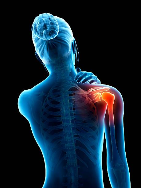
- Anatomy of the shoulder
- Types of injuries
- MRI and repairability criteria
- Arthroscopic repair
- Suture techniques
- Arthroscopic repair
- Simple suture
- Double row
- Post-operative physiotherapy
- Avoid waiting too long before surgery
- MRI-arthrography is the examination of choice
- Rigorous physiotherapeutic follow-up after suture
Information
Video type:
Anatomy:
Surgery:
Thematic:
Penetrating lesions: definition, issues and diagnosis
A penetrating lesion involves the entire thickness of the tendon and interrupts contact with the bone. Clinical findings suggest pain in elevation and rotation, sometimes at night, with weakness. MRI arthrography specifies tendon retraction and muscle trophicity, key elements of repairability.
Identifying these criteria early prevents the progression to difficult conditions, particularly in active patients.
Surgical indications: act at the right time
Waiting too long exposes the patient to progression towards irreparability. MRI arthrography has a limited decision-making value; after six months, a new examination may be necessary if the clinical picture changes.
The indication is based on functional impairment, age, risk factors and muscle quality.
A penetrating lesion will not heal spontaneously.
Arthroscopic repair: principles and steps
The repair is performed arthroscopically through small incisions. After bone preparation, anchors are placed, the threads are passed through the tendon and then reapplied to the greater tuberosity. The technique is tissue-friendly and allows for standardized care.
Depending on the extent and the tendons involved, the location and number of anchors are adapted.
Single or double row fixing: contact surface and stability
Double row increases the tendon-bone contact area and can improve biomechanical stability. It is not essential in all cases, but is readily incorporated for larger defects.
The objective remains the restoration of a solid anchoring and harmonious kinematics.
Nature needs three months for the tendon to bond to the bone.
Biological time and rehabilitation: protect to recover better
The first six weeks are devoted to passive mobility and pain control. Any premature loading compromises tendon-bone adhesion. This is followed by progressive activation and targeted strengthening, within a clear educational framework.
Close coordination between surgeon, patient and physiotherapist determines the result.
Results and prognostic factors
Satisfaction rates are high, with sustained improvement in pain and function. Lesion size, retraction, multi-tendon involvement, and preoperative cortisone injections affect healing.
Repairing in time limits the risk of secondary osteoarthritis by restoring more physiological glenohumeral mechanics.
Pathologies treated at the center
Hallux Limitus
Functional
Your pain has a cause.The balance sheet allows us to understand it.
- Gait analysis
- Posture Assessment
- Guidance on the right treatment
- Study of plantar supports and supports
- Detection of compensations
- Pain–movement correlation
The functional assessment allows us to understand how a joint or postural imbalance can trigger or perpetuate pain. Very often, imaging is normal, but movement is disturbed. By analyzing gait, weight-bearing patterns, or posture, we identify the weak links in the chain and guide targeted treatment adapted to the patient's actual mechanics.