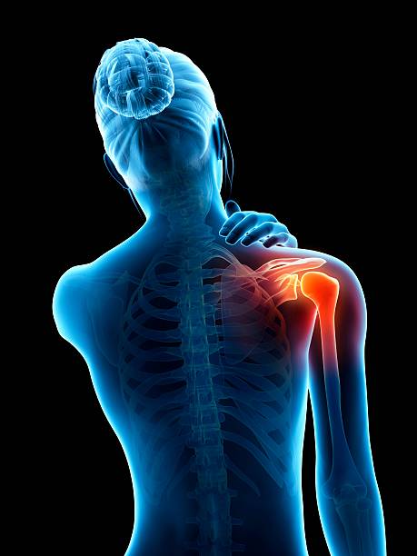
Arthroscopic rotator cuff repair: indications, technical procedures and follow-up
Continuing education on rotator cuff injuries with a dual intervention: clinical diagnosis and imaging, followed by a commented surgical demonstration. This video provides a comprehensive overview of the treatment pathway, from clinical tests to surgical indications, and discusses the role of conservative or prosthetic treatments depending on the case.
Doctors
Topics
Treatments
Advice
- Dr. Steve Brenn; Dr Ludovic Glanz
- Anatomy of the cuff
- Clinical tests
- Diagnostic imaging
- Repairability criteria
- Surgical treatment
- Arthroscopic repair
- Thenodesis
- Infiltration
- Reverse prosthesis
- Do not delay repair after traumatic rupture
- Importance of pre- and post-operative physiotherapy
- MRI with arthrography as the examination of choice
- Thenodesis preferable to thenotomy in most cases
- Double row improves repair stability
Information
Video type:
Anatomy:
Thematic:
From diagnosis to action: a structured trajectory
The management of a rotator cuff injury begins with a rigorous clinical examination (Job's tests, countered external rotation, belly press) combined with appropriate imaging. This process allows the tear to be qualified, associated injuries to be identified and the surgical plan to be anticipated if necessary.
The therapeutic strategy is graduated: patient information, targeted physiotherapy, then surgery when pain persists, function is limited and anatomical criteria allow for reliable repair.
Imaging and decision criteria
MRI arthrography is preferred for characterizing the extent of the rupture, retraction, fatty infiltration, and cartilage condition. These elements guide the risk-benefit balance between conservative treatment, repair, or alternative prosthetics. The existence of a short delay after a traumatic rupture in an active patient is a strong argument for earlier repair.
Corticosteroid injections should be used with caution before planned surgery, due to their impact on healing and the risk of infection.
MRI-arthrography is the examination of choice for diagnosing rotator cuff lesions.
Arthroscopic procedure: freshening, anchoring and double row
Arthroscopic repair aims to reapply the tendon to its bony imprint after careful preparation of the site (freshening of the footprint). Resorbable or metallic inks, connected to strong sutures, ensure fixation. The double-row configuration increases the tendon-bone contact surface and mechanical stability, factors correlated with the quality of healing and the maintenance of the result over time.
An “on-demand” acromioplasty can be performed to reduce residual conflict when the morphology of the acromion justifies it.
Role of the biceps: thenodesis and alternatives
Anterior pain related to the long head of the biceps is common in rotator cuff injuries. Depending on age, activity, and associated injury, thenodesis (reattachment in the bicipital groove) is often preferred, providing pain control and good functional outcome. Tenotomy may be discussed in selected profiles, at the cost of a risk of Popeye sign and aesthetic variability.
The choice fits into the overall project: repair what can be repaired, treat pain and preserve function.
The double row increases the contact surface and stability of the repair.
Rehabilitation: phases and objectives
Rehabilitation follows clearly defined stages: relative immobilization and early passive mobilizations to limit stiffness, then active assisted after six weeks, and progressive strengthening from the third month. This chronology respects the tendon-bone healing time and determines the strength of the repair.
The objectives are the recovery of range of motion, dynamic refocusing of the shoulder and the safe resumption of professional and sporting activities.
What to remember for a lasting shoulder
An accurate diagnosis, an indication based on objective criteria, and a rigorous arthroscopic technique allow for long-lasting results. Education regarding biological timescales and adherence to rehabilitation are crucial for preserving repair and avoiding recurrence.
The purpose is functional: to reduce pain, restore strength and enable confident use of the shoulder.
Pathologies treated at the center
Hallux Limitus
Functional
Your pain has a cause.The balance sheet allows us to understand it.
- Gait analysis
- Posture Assessment
- Guidance on the right treatment
- Study of plantar supports and supports
- Detection of compensations
- Pain–movement correlation
The functional assessment allows us to understand how a joint or postural imbalance can trigger or perpetuate pain. Very often, imaging is normal, but movement is disturbed. By analyzing gait, weight-bearing patterns, or posture, we identify the weak links in the chain and guide targeted treatment adapted to the patient's actual mechanics.