Anatomy, biomechanics and understanding of meniscal injuries
This video reviews the anatomical and biomechanical foundations of the meniscus to better understand the origin of lesions. It shows how remote anomalies (hallux valgus, femoral rotation, etc.) can affect the knee. This provides valuable insight for adapting diagnosis and treatment.
Doctors
Topics
Treatments
Advice
- Dr Jacques Vallotton
- Anatomy of the menisci
- Joint biomechanics
- Extrinsic factors
- Foot-knee link
- Prevention
- Prevention
- Clinical analysis
- Therapeutic education
- Take into account the overall morphotype
- Correlate MRI and clinical
- Preserve the lateral meniscus as much as possible
Information
Video type:
Anatomy:
Surgery:
Anatomy and functional role: why the meniscus is essential
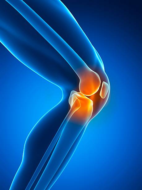
The meniscus, a semilunar fibrocartilaginous structure, increases the femorotibial contact surface, stabilizes the joint and absorbs loads. Its mobility allows congruence to be adapted to each flexion angle, particularly in the lateral compartment where the popliteal hiatus makes the capsule discontinuous. This dynamic protects the cartilage and contributes to knee proprioception.
Meniscus damage disrupts this protective mechanism. Stress refocuses, increases, and accelerates cartilage wear. Understanding this function is essential for interpreting a lesion and choosing the most appropriate treatment.
Kinematics, kinetics and repeated overload
Meniscal kinematics describes the movement of the tissue during flexion-extension; the contact point slides backward in flexion, especially laterally, imposing a meniscal recoil. Kinetics refers to the local constraints linked to the gesture (support, strike), which particularly stress the middle segment of the medial meniscus. Through micro-impactions, a segment becomes rigid, shifting the constraints towards the horns and promoting tears.
Repeated overloading of a compartment can lead to meniscal cysts or bone indentations, indicating mechanical imbalance. Reducing these overloads is a major therapeutic goal.
The menisci are mobile and essential at every angle of flexion.
Beyond the knee: extrinsic factors and the functional chain
The knee is part of a chain. A functional hallux limitus can project the trunk forward, increase the flexion moment, and create a medial collapse with each step. Femoral antetorsion, tibial torsion, varus/valgus, and dysmetria also influence the load. Over millions of annual cycles, these parameters shape the stress trajectory and explain certain so-called "wear and tear" injuries.
From then on, the clinical assessment integrates gait, hip mobility and foot statics. Targeted correction may be sufficient to normalize mechanics and calm symptoms.
MRI and anatomical variants: avoiding the pitfalls
MRI interpretation requires contextual reading. The Wiesberg ligament, involved in the posterior horn of the lateral meniscus, can simulate a tear. The lateral compartment, more mobile and less fixed to the capsule, generates singular images. Clinical relevance therefore takes precedence over the radiological signal alone.
Correlating imaging with clinical tests, mechanical pain and functional impact avoids untimely actions on abnormalities without symptomatic significance.
A meniscal injury must always be placed in its overall context.
Support: Priority to function and preservation
Initial treatment focuses on rehabilitation, load management, and correction of extrinsic factors. When mechanical pain persists and annular integrity is compromised (radial tear, root lesion), repair is discussed. Preservation of the lateral meniscal tissue, the pillar of stability of the external compartment, is a priority.
In some cases, correction of the axis (osteotomy) accompanies the repair to permanently rebalance the loads.
Perspective for the patient
Care focused on individual mechanics often makes it possible to avoid surgery. If surgery is necessary, the goal is to restore the meniscal ring and knee kinematics, which are essential for a good functional prognosis. Education on daily and sporting activities helps prevent recurrence and protect cartilage.
The essential message remains constant: understand, correct, preserve.
Pathologies treated at the center
Hallux Limitus
Functional
Your pain has a cause.The balance sheet allows us to understand it.
- Gait analysis
- Posture Assessment
- Guidance on the right treatment
- Study of plantar supports and supports
- Detection of compensations
- Pain–movement correlation
The functional assessment allows us to understand how a joint or postural imbalance can trigger or perpetuate pain. Very often, imaging is normal, but movement is disturbed. By analyzing gait, weight-bearing patterns, or posture, we identify the weak links in the chain and guide targeted treatment adapted to the patient's actual mechanics.


