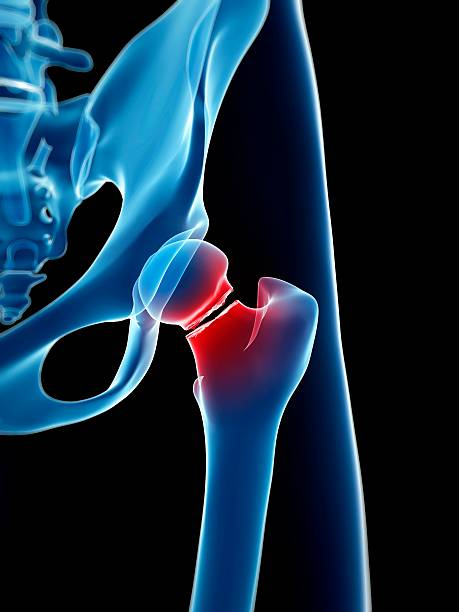
Differential diagnosis of hip pain
In this presentation, Dr. Vallotton explores the biomechanical origins of hip pain and emphasizes the importance of differential diagnosis. He discusses anatomical variations, body type analysis, and clinical tests to perform, particularly in athletes or adolescents with atypical pain.
Doctors
Topics
Treatments
Advice
- Dr Jacques Vallotton
- Biomechanics of the hip
- Comparative anatomy
- Morphotype analysis
- Clinical tests
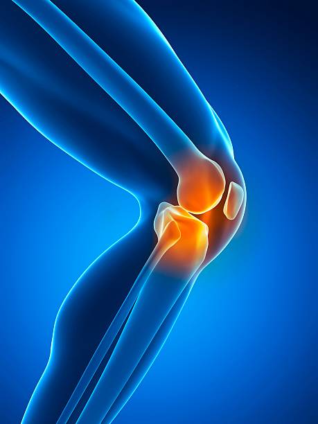
- Link between hip and knee pain
- Morphotype analysis
- Clinical examination
- Radiological assessment
- Systematically examine the hip in case of knee pain
- Analyze pelvic rotation
- Considering adolescent growth and sport
Information
Video type:
Anatomy:
Surgery:
Hip pain: remember the biomechanics to better understand
The hip is a powerful ball-and-socket joint whose anatomy varies enormously from one individual to another. The evolution towards bipedalism has shaped the pelvis, femoral valgus, and locomotor chain, explaining very different morphotypes. This variability conditions load distribution and exercise tolerance.
Understanding these bases sheds light on the origin of the pain and prepares the clinical examination: some hips are very "hollow" at the neck, others more "bulging", or even contrasting acetabular covers influence the areas of conflict.
Anatomical variations and effects of activity
Head-neck angle, offset, acetabulum coverage, and femoral version vary widely. In adolescent athletes, repeated stresses can remodel the head-neck junction and promote femoroacetabular impingement. Tibial torsion and pelvic orientation complete the morphological picture.
The analysis must be bilateral and dynamic in order to assess asymmetries and their functional consequences.
Knee pain may be a sign of a hip problem.
Morphotype, gait and hip-hindfoot synchronism
The lower limb combines two "ball and socket" joints - hip and subtalar - which are synchronous with walking. Foot pronation is accompanied by tibial rotation and knee adaptations, while pelvic tilt modifies muscle play.
Observing gait is as essential as static examination: step angle, rotations and frontal stability provide information on actual load constraints.
Clinical examination: look at the pelvis above the femoral head
Assessment in the ventral decubitus position, with the capsule extended, specifies internal/external rotations. The gluteus medius stabilizes the pelvis in single-leg support; its failure causes tilting and excessively stresses the piriformis, which is often contracted by compensation.
This change of frame of reference – following the movement of the pelvis rather than that of the femur – helps to understand strength imbalances and lameness.
Gait analysis shows us the overall functioning of the individual.
Differential diagnosis and rational imaging
Differential diagnosis is based on the location of the pain (groin crease, buttock, knee), triggering factors, palpation, and functional tests. In adolescents, epiphysiolysis should be considered. Imaging (targeted X-rays, sequential sections) specifies torsions and covers when necessary.
This approach avoids therapeutic dead ends and guides towards the right treatment.
Support: Restore balance, preserve function
The strategy combines education, muscle rebalancing, correction of mechanical factors and, if necessary, targeted movements. The goal is to restore harmonious movement and avoid painful compensations in the locomotor chain.
Any knee pain prompts examination of the hip; a comprehensive and graduated approach remains the best guarantee of lasting functional return.
Pathologies treated at the center
Hallux Limitus
Functional
Your pain has a cause.The balance sheet allows us to understand it.
- Gait analysis
- Posture Assessment
- Guidance on the right treatment
- Study of plantar supports and supports
- Detection of compensations
- Pain–movement correlation
The functional assessment allows us to understand how a joint or postural imbalance can trigger or perpetuate pain. Very often, imaging is normal, but movement is disturbed. By analyzing gait, weight-bearing patterns, or posture, we identify the weak links in the chain and guide targeted treatment adapted to the patient's actual mechanics.