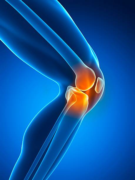
MRI and meniscal injuries after an ACL sprain: what do you need to know?
This presentation explores in detail meniscal injuries associated with ACL sprains, focusing on MRI images and the recognition of longitudinal and radiating tears. It addresses the anatomical specificities of the medial and lateral meniscus, the importance of imaging timing, and the clinical implications of these under-recognized injuries. This content is essential for improving post-traumatic knee diagnosis.
Doctors
Topics
Treatments
Advice
- Dr. Henri Guérini
- Radiological anatomy of the meniscus
- Posterior longitudinal lesions
- Meniscofemoral ligaments
- Acute phase MRI
- Ramp-type cracks
- MRI
- Arthroscanner
- Post-traumatic monitoring
- Longitudinal lesions are often located behind the lateral meniscus
- Humphrey and Friedberg ligaments are key MRI landmarks
- Early post-sprain MRI improves injury detection
- Some lesions heal spontaneously
- Ramp lesions are often underdiagnosed
Information
Video type:
Anatomy:
Surgery:
Thematic:
After an ACL sprain: Why an MRI matters
ACL sprains are often accompanied by meniscal injuries, which are sometimes unrecognized. MRI, performed at the right time, helps identify these injuries and distinguish between injuries requiring simple monitoring and those that expose them to mechanical instability. This step improves therapeutic dialogue and the planning of rehabilitation or possible surgery.
The challenge is twofold: to better understand the cause of pain and to prevent blockages or cartilage degradation.
What MRI looks for
Posterior longitudinal fissures and so-called "ramp" lesions are common after ACL sprains. The lateral meniscus is often affected in the acute phase; some internal lesions appear later. Imaging also identifies radiated lesions of the posterior horns and capsulomeniscal detachments, the management of which differs depending on the area and stability.
Accurately identifying the type of injury guides treatment and loading instructions.
We should not focus only on the ACL and forget about meniscal lesions in MRI.
The right timing for the MRI
Contrary to popular belief, performing an MRI close to the accident can help: edema and effusion highlight tear lines and detachments that are difficult to see later. This diagnostic window improves the sensitivity of the examination and prevents unstable lesions from being missed.
When MRI is late or ambiguous, targeted monitoring can be discussed depending on the clinical course.
Therapeutic consequences
Many peripheral lesions heal with appropriate protection and rehabilitation; others, which are unstable, benefit from suturing to preserve the anteroposterior and rotational stability of the knee. Preserving the meniscus, when possible, protects the joint in the long term.
The decision is based on location, vascularity, stability and functional goals.
Performing an MRI close to the accident allows for better visualization of the lesions thanks to the edema.
What the patient should remember
MRI is a tool, not an end in itself. When used properly, it clarifies the situation, avoids unnecessary procedures, and ensures a safe return to work. It is always integrated into the clinical examination and rehabilitation plan, ensuring a return to function in good conditions.
Perspective
A stable knee and preserved menisci are essential for joint performance and longevity. Careful reading of MRI scans at the right time directly contributes to this.
Pathologies treated at the center
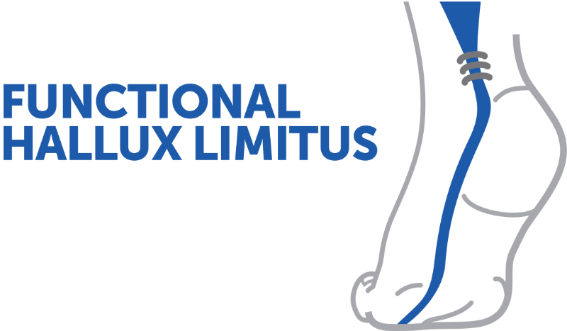
Hallux Limitus
Functional
Your pain has a cause.The balance sheet allows us to understand it.
- Gait analysis
- Posture Assessment
- Guidance on the right treatment
- Study of plantar supports and supports
- Detection of compensations
- Pain–movement correlation
The functional assessment allows us to understand how a joint or postural imbalance can trigger or perpetuate pain. Very often, imaging is normal, but movement is disturbed. By analyzing gait, weight-bearing patterns, or posture, we identify the weak links in the chain and guide targeted treatment adapted to the patient's actual mechanics.


