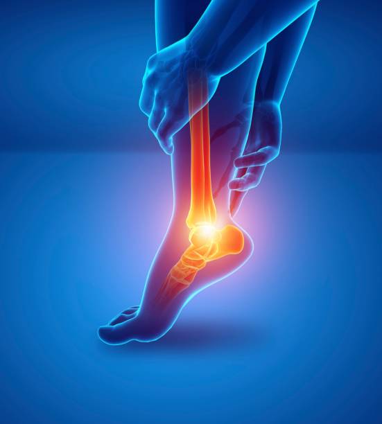
MRI imaging of the rotator cuff tendons: value of ABER sequences
Presentation of the different imaging techniques (MRI, MRI-arthrography, ABER position) used to assess rotator cuff tendon injuries. The intervention explains how to differentiate the types of tears (non-penetrating, penetrating), the locations of the injuries, as well as the criteria to consider when directing surgical or non-surgical treatment.
Doctors
Topics
Treatments
Advice
- Dr. Delphine Richarme
- Imaging techniques
- MRI sequences
- Types of injuries
- ABER position
- Preoperative evaluation
- Imaging assessment
- Arthro-MRI
- Simple MRI
- Arthro-CT
- MRI-arthrography is the gold standard for deep tears.
- ABER position improves detection of deep partial lesions
- the type of lesion guides surgical treatment
Information
Video type:
Anatomy:
Surgery:
Thematic:
Why rotator cuff imaging is critical
Shoulder pain related to the rotator cuff is common and can disrupt strength, mobility, and sleep. Imaging can help clarify the nature of the injury, assess its severity, and guide treatment. Distinguishing tendinopathy from a partial or full-thickness tear determines management, as does the exact location of the lesion (bursal, articular, or intratendinous surface). The choice of modality is not trivial: each technique provides complementary information, useful for deciding on targeted rehabilitation or surgery. In this context, the radiologist describes not only the affected tendon (often supraspinatus and infraspinatus), but also the condition of the long biceps and subscapularis, elements closely associated with glenohumeral stability. The goal is to provide a readable, decision-oriented report that links the image to the therapeutic procedure and recovery schedule.
Thus, the imaging examination is not limited to "seeing a tear": it serves to quantify the damage, specify its location, identify indirect signs (effusion, bursitis) and indicate the factors which influence repairability.
MRI, MRI-arthro and CT-arthro: complementary indications
Three approaches are used: conventional MRI, MRI-arthrography with intra-articular injection of gadolinium, and CT-arthrography (rarer in Switzerland). Simple, non-invasive MRI characterizes the tendons and bursae in a neutral position. MRI-arthrography, the reference examination for deep lesions, highlights the diffusion of the contrast agent through a full-thickness tear and improves the detection of damage to the articular surface. CT-arthrography remains a relevant alternative in patients contraindicated to MRI (claustrophobia, highly artifactogenic material, allergy to gadolinium). The report specifies the affected tendon(s), the lesion topography and any associated damage to the long biceps. This cross-reading, correlated with the clinical picture, allows conservative treatment to be adapted or tendon repair to be planned when justified.
At this stage, it should be remembered that the indication for examination depends on the clinical context; the objective is to obtain a reliable map that can be used by the healthcare team.
MRI-arthrography is truly the most effective examination for evaluating the rotator cuff tendons.
Understanding the types and locations of tears
A rotator cuff injury can be partial (bursal, articular, or interstitial) or transfixing. On MRI arthrography, diffusion of contrast in the subacromial bursa confirms transfixion. Identifying the affected area guides the strategy: injury to the articular surface often reflects different mechanical constraints than a bursal injury. The radiologist also analyzes tendon thickness; a normal tendon measures approximately 10 to 12 mm, which helps estimate substance loss and chronicity. Subscapularis injuries, sometimes responsible for instability of the long biceps, are better assessed on axial slices. These elements, added to the context (traumatic vs. degenerative), make it possible to anticipate the complexity of a possible repair and inform the patient about realistic recovery goals.
Finally, the presence of bursitis, effusion or alterations of the long biceps contributes to the overall understanding of the picture.
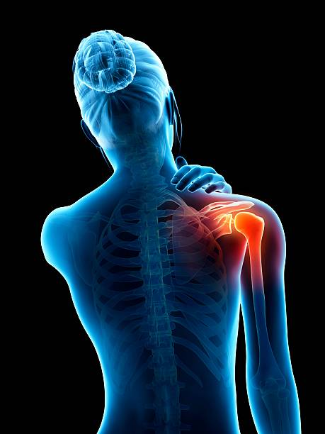
The specific role of sequences in ABER position
The ABER position (abduction and external rotation) relaxes the cuff and modifies the relationship between the tendon and the humeral head. It improves the visualization of deep lesions, particularly on the articular surface, and can reveal fissures that are not evident in the neutral position. The clinical interest is twofold: to refine the diagnosis when doubt persists after standard sequences, and to better estimate the true extent of a partial lesion. In this context, ABER complements MRI arthrography and increases sensitivity for fine tears of the supraspinatus. Its interpretation requires systematic reading of the section planes (coronal, sagittal and axial) in order not to overlook subscapularis damage or long biceps instability.
Thus, ABER is not systematic, but becomes decisive when the clinical picture suggests pain on elevation or external rotation with standard imaging showing little contribution.
The ABER position allows the tendons to relax and deep lesions to be better visualized.
What a useful report for the surgeon should contain
A usable report specifies: the affected tendon(s), the location (bursal, articular, interstitial), the depth and length of the crack, any transfixion, the condition of the long biceps and subscapularis. Tendon thickness (reference 10–12 mm for a healthy tendon) and the overall quality of the tissues provide guidance on chronicity. Sagittal and axial analysis looks for associated signs (bursitis, synovitis, secondary impingement). This information, correlated with functional age and activity level, allows for discussion of rehabilitation, rational infiltration or arthroscopic repair. In this context, the objective is to avoid delays in treatment while selecting the relevant indications.
This structured approach secures the decision shared with the patient and optimizes surgical planning when necessary.
Message to the patient: an examination to inform the decision
Imaging is not an end in itself; it serves to understand the origin of pain and choose the best strategy. Depending on the context, a simple MRI may be sufficient; in other situations, MRI arthrography and the ABER position offer more detailed answers, particularly for deep lesions. It is normal for an examination to be proposed even if the pain varies from day to day: the objective is to map the lesion and evaluate the associated structures. However, the indication for an operation is not based solely on the image; it combines symptoms, activity, functional objectives, and tissue quality. Thus, a clear imaging result facilitates personalized and realistic treatment, from rehabilitation to possible repair.
Pathologies treated at the center
Hallux Limitus
Functional
Your pain has a cause.The balance sheet allows us to understand it.
- Gait analysis
- Posture Assessment
- Guidance on the right treatment
- Study of plantar supports and supports
- Detection of compensations
- Pain–movement correlation
The functional assessment allows us to understand how a joint or postural imbalance can trigger or perpetuate pain. Very often, imaging is normal, but movement is disturbed. By analyzing gait, weight-bearing patterns, or posture, we identify the weak links in the chain and guide targeted treatment adapted to the patient's actual mechanics.