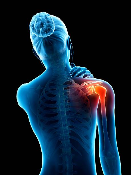
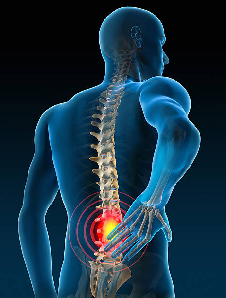
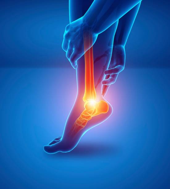
Postpartum pelvic pain: a comprehensive and interdisciplinary approach
Through a complex clinical case, Dr. Vallotton and his colleagues discuss a patient presenting with pelvic and lower back pain after multiple deliveries. The analysis highlights the importance of a comprehensive approach combining rehabilitation, orthotics, and interdisciplinary support to restore function and provide lasting relief.
Doctors
Topics
Treatments
Advice
- Dr Jacques Vallotton
- Postpartum clinical case
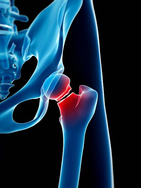
- History and pelvic instability
- Biomechanical approach
- Treatments considered
- Perineal rehabilitation
- Muscle rehabilitation
- Plantar orthoses
- Infiltrations
- PRP
- Multidisciplinary approach
- progressive rehabilitation
- overall functional assessment
- muscle stabilization
- avoid early racing
- role of footbeds
Information
Video type:
Anatomy:
Surgery:
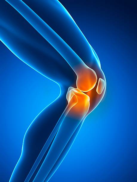
Patellar instability: putting the knee in context
Patellar instability is not just anterior knee pain. It results from interactions between bone morphology, muscle balance, femoro-tibial rotations, and quality of support. From the outset, the clinical assessment is integrated into a global reading of the lower limb and dynamic posture.
This perspective guides the choice of examinations and treatments, from targeted rehabilitation to surgical solutions when necessary.
Clinical testing: apprehension, translation and tilt
Three tests dominate: the engagement sign, the apprehension test and the lateral translation, which provide information on active and passive stability. Patellar tilt, spontaneous or iatrogenic, completes the examination and alerts to excessive lateral tension.
The reproducibility of these maneuvers makes them reliable benchmarks for objectifying instability and monitoring the effect of treatment.
Engagement, apprehension and translation are the three most representative tests.
Reasoned imaging: from standard image to MRI
The standard radiological assessment requires a strict profile and a 30° axial angle. The CT scan specifies torsions and allows for an estimation of the TAGT, a useful measurement but to be interpreted according to the morphotype and the recurvatum. The MRI provides a detailed cartilaginous and ligamentous reading, identifies free bodies and characterizes the trochlear height.
Beyond the figures, it is clinical-imaging coherence that guides the therapeutic plan.
Role of walking and support: the fifth dimension
The gait assessment and podiatric analysis objectify the supination-pronation sequences, pelvic tilts and trunk adaptations. These parameters condition the patellar trajectory and explain subtle instabilities such as episodes of subluxation.
Integrating this data makes it possible to adjust rehabilitation (sheathing, lateral chain, postural control) and better select surgical indications.
The walking assessment will become an essential examination in the future.
Treatments: reasoned progression and personalization
The first line of treatment combines neuromuscular rehabilitation, weight-bearing optimization, and pain management. When trochlear dysplasia, high patella, or lateralization are severe, MPFL reconstruction, trochlear procedures, or tuberosity medialization are discussed.
The choice is based on alignment of evidence: clinical, imaging, and response to conservative trials.
Functional objective: stability, confidence and return to activities
Success is measured by regained stability, the disappearance of apprehension, and the resumption of activities without fear of slipping. Careful monitoring consolidates the results through adjustments to strengthening and movement technique.
The therapeutic trajectory remains individualized, with a clear direction: preserve the cartilage and secure patellar kinematics.
Pathologies treated at the center
Hallux Limitus
Functional
Your pain has a cause.The balance sheet allows us to understand it.
- Gait analysis
- Posture Assessment
- Guidance on the right treatment
- Study of plantar supports and supports
- Detection of compensations
- Pain–movement correlation
The functional assessment allows us to understand how a joint or postural imbalance can trigger or perpetuate pain. Very often, imaging is normal, but movement is disturbed. By analyzing gait, weight-bearing patterns, or posture, we identify the weak links in the chain and guide targeted treatment adapted to the patient's actual mechanics.