The Evolution of the Human Foot: A Paleontological Perspective
In this original presentation, Dr. Vallotton explores the evolution of the human foot through the prism of paleontology, highlighting anatomical specificities, locomotor adaptations and the implications for certain current pathologies such as hallux valgus.
Doctors
Topics
Treatments
Advice
- Dr Jacques Vallotton
- Evolution of vertebrates
- Specificities of the human foot
- Human-ape differences
- Archaic reflexes
- Biomechanical consequences
- Biomechanical analysis
- Orthoses
- Evolutionary thinking
- Paleontological origin of the human foot
- Importance of MP1 in locomotion
- Influence of evolution on current pathologies
- Link between bone shape and posture
Information
Video type:
Anatomy:
Surgery:
Thematic:
Why the human foot still poses questions
The foot is a highly complex locomotor organ, the result of a long and non-linear evolutionary history. Its current shape reflects adaptive choices that are not identical to those of other primates. Understanding these trajectories sheds light on walking, running, and certain forefoot pathologies observed today.
From this perspective, paleontological reading provides benchmarks: the appearance of the first primates, occasional bipedalism of australopithecines, and the progressive acquisition of mechanics conducive to endurance. These stages shaped an architecture adapted to prolonged support and economy of effort.
Paleontological landmarks: from mangroves to plains
Evolutionary hypotheses suggest a proto-hominid ancestor capable of evolving in mixed aquatic and terrestrial environments. Over time, the verticalization of the talus above the calcaneus, the elongation of the levers, and group organization favored endurance running. These adaptations modified the use of the foot and the synchronization of the proximal chains.
The fossil discoveries, although fragmentary, show variations in footprints, the position of the big toe, and the shape of the calcaneus as markers of evolution. They shed light on a path where environmental constraints, posture, and movement strategies coexist.
The human foot is a constantly evolving organ.
Comparing man and ape: two propulsion mechanisms
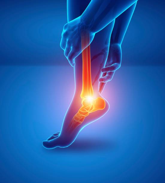
In humans, dorsiflexion at the end of stance occurs primarily at the first ray metatarsophalangeal joint, promoting efficient propulsion. In monkeys, flexion is more organized at the midtarsal joint, making the foot more prehensile but less efficient for prolonged walking. This difference explains distinct motor strategies and characteristic podiatric signatures.
Thus, where man seeks dynamic stability with a first guiding ray, the monkey values adaptation and grip. The contrast highlights the role of the big toe in the orientation of the forefoot and the continuity of support.
The “coxa pedis”: a hinge for stability
The talocalcaneonavicular joint functions like a true hip of the foot. Its rotational action coordinates the pronation-supination transition and synchronizes the alignment of the overlying segments. A blockage or shift in this rhythm is reflected down to the knee and hip, with sometimes costly postural adaptations.
This functional reading reminds us that walking is an orchestration: a local restriction can shift constraints elsewhere. Clinical observation must therefore integrate the entire chain, and not just the plantar footprint.
We have probably retained a very primitive foot.
Morphological variations: diversity and constants
Foot morphology shows significant variations between individuals: width of the calcaneus, arches, orientation of the first ray, and depth of the retrotalar groove. These differences, the result of habitus and adaptations, are expressed in the footprint and walking kinematics.
Despite this diversity, certain constants remain: the leading role of the hallux in propulsion, alternating pronation and supination, and the search for dynamic balance. The analysis must distinguish between what is an anatomical variant and what disrupts function.
Clinical implications: shedding light on certain pathologies
Evolutionary understanding helps interpret the emergence of forefoot disorders, including hallux valgus and certain rigidus. Rapid height and weight growth and modern constraints can exceed adaptive capacities, creating mechanical conflicts. Linking form to function allows for more coherent management strategies.
In practice, the examination combines observation of gait, targeted mobility tests and reading of support. This global view places the foot in the continuity of the lower limb and posture.
Pathologies treated at the center
Hallux Limitus
Functional
Your pain has a cause.The balance sheet allows us to understand it.
- Gait analysis
- Posture Assessment
- Guidance on the right treatment
- Study of plantar supports and supports
- Detection of compensations
- Pain–movement correlation
The functional assessment allows us to understand how a joint or postural imbalance can trigger or perpetuate pain. Very often, imaging is normal, but movement is disturbed. By analyzing gait, weight-bearing patterns, or posture, we identify the weak links in the chain and guide targeted treatment adapted to the patient's actual mechanics.