Understanding hip osteoarthritis and its treatments
This video explains hip osteoarthritis, its causes, symptoms, and treatments. It details conservative solutions such as physiotherapy, injections, and weight loss, followed by surgical options such as hip replacements, with an overview of materials, innovations, and rehabilitation.
Doctors
Topics
Treatments
Advice
- Prof. Brigitte Jolles-Haeberli
- Definition of osteoarthritis
- Causes and symptoms
- Clinical and radiological diagnosis
- Conservative treatments
- Total hip replacement
- Conservative treatment
- Total hip replacement
- Hyaluronic acid infiltration
- Watch for early symptoms of groin pain
- Maintain hip mobility
- Reduce weight to protect the joint
- Consult in case of lameness
- Respect post-operative rehabilitation
Information
Video type:
Anatomy:
Thematic:
Understanding hip osteoarthritis
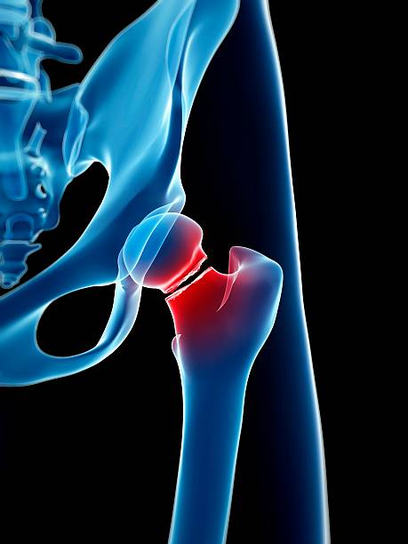
Hip osteoarthritis is a progressive wear of the cartilage covering the femoral head and acetabulum. When this smooth surface loses its integrity, sliding becomes irregular and painful, causing discomfort when walking and performing daily activities. The joint then seeks to compensate by peripheral bone proliferation, at the cost of loss of mobility and muscle fatigue. In this context, early diagnosis and clear information on treatment options make it possible to anticipate the necessary lifestyle adjustments.
This pathology affects a variety of profiles. It can be part of physiological aging, result from a history of trauma, or be associated with rheumatic diseases. In younger individuals, a morphological abnormality (dysplasia) accelerates wear. The initial goal is to preserve function by maintaining the most fluid gait possible, as cartilage is nourished by joint fluid through movement. A progressive strategy—guided exercises, weight control, and reasoned medical treatment—is the first step.
Symptoms and risk factors
The pain typically occurs in the groin crease, sometimes radiating toward the thigh. It worsens with weight bearing, when climbing stairs, or when getting up from a prolonged sitting position. Limping may occur with weakness of the gluteal muscles and a marked decrease in internal rotation. Getting dressed, putting on shoes, or cutting nails becomes difficult. These symptoms reflect a loss of joint congruity and bone-on-bone friction in the advanced stages.
Several factors increase the risk: heredity, sports with repeated impacts, being overweight (each additional kilo increases the constraints), certain rheumatic conditions, old traumatic sequelae and, more rarely, fragile vascularization of the femoral head favoring subchondral necrosis. Identifying these elements guides prevention (weight reduction, adaptation of activities) and guides therapeutic discussion at the right time.
Once the cartilage is worn, the femoral head can no longer slide properly.
Clinical and radiological assessment
The examination begins with gait analysis: looking for limping, pelvic tilt, and spinal compensation. Lying down, testing often reveals pain on internal rotation, with relatively preserved external rotation. Simple maneuvers (piston, rolling) objectify the loading pain. A rapid neurological and vascular examination can rule out associated diagnoses.
Standard X-rays (frontal pelvis, profiles) are usually sufficient to confirm the diagnosis: joint narrowing, osteophytes, subchondral geodes and sclerosis are characteristic. 3D planning by scanner is useful preoperatively to simulate the positioning of implants, assess stress areas and optimize the balance between stability and mobility after surgery.
Conservative treatments and healthy lifestyle
The first step aims to improve joint glide and muscle guidance. Physiotherapy restores range of motion, maintains gait, and reduces pain by promoting the diffusion of joint fluid. Weight reduction reduces constraints. Depending on the context, progressive analgesic treatment (paracetamol, anti-inflammatories, and then, more rarely, opioids) may be necessary. In young patients wishing to postpone surgery, viscosupplementation with hyaluronic acid may be discussed.
The goal is to achieve acceptable functioning for daily life, delay the need for surgery, and preserve muscle mass. Regular support helps adapt activity and identify when medical treatment becomes insufficient.
The goal of surgery is to regain painless support and good mobility.
Total hip replacement: options and materials
When pain limits walking and basic movements despite well-conducted treatment, total hip replacement becomes appropriate. The anterior or posterior approaches are chosen according to the team's habits and anatomy. Implants can be cemented (immediate full weight-bearing, useful in cases of osteoporosis) or uncemented, with a hydroxyapatite coating promoting osseointegration (transient partial weight-bearing).
The friction torque determines long-term wear: ceramic–highly cross-linked polyethylene, metal–polyethylene, ceramic–ceramic, or metallized ceramic (zirconium). Dual-mobility heads increase the arc of motion and reduce the risk of dislocation in certain active profiles. 3D planning, custom guides, and, in some centers, robotic assistance contribute to more precise reconstruction.
Rehabilitation and prospects
Mobilization begins early: walking with canes, strengthening the gluteal and quadriceps muscles, and learning to climb stairs from the first days. Support is adapted to the type of fixation until the 4–6 week check-up, then the goal is to achieve smooth walking without canes and gradually resume non-impact leisure activities. Satisfaction is high when pain is controlled and function restored.
Current advances include systematic 3D planning, optimization of friction torques, and implant customization. Success lies in the combination of timely indication, precise execution, and rigorous rehabilitation.
Pathologies treated at the center
Hallux Limitus
Functional
Your pain has a cause.The balance sheet allows us to understand it.
- Gait analysis
- Posture Assessment
- Guidance on the right treatment
- Study of plantar supports and supports
- Detection of compensations
- Pain–movement correlation
The functional assessment allows us to understand how a joint or postural imbalance can trigger or perpetuate pain. Very often, imaging is normal, but movement is disturbed. By analyzing gait, weight-bearing patterns, or posture, we identify the weak links in the chain and guide targeted treatment adapted to the patient's actual mechanics.