Understanding shoulder osteoarthritis and its treatments
This educational video presents shoulder osteoarthritis: the joints involved, typical symptoms (pain, loss of mobility, inflammation), and conservative or surgical treatment options. It also explains the difference between anatomic and reverse prosthesis, in simple terms accessible to the general public.
Doctors
Topics
Treatments
Advice
- Dr. Ludovic Glanz
- Definition of osteoarthritis
- Symptoms and pain
- Conservative treatment
- Anatomical and reverse prosthesis
- Role of the deltoid
- Conservative treatment
- Anatomical prosthesis
- Reverse prosthesis
Information
Video type:
Anatomy:
Surgery:
Thematic:
Understanding Shoulder Osteoarthritis: Anatomy and Mechanisms
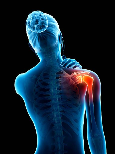
The shoulder is not a single joint but a functional whole where the scapulothoracic, sternoclavicular, acromioclavicular and especially the glenohumeral work together. In glenohumeral osteoarthritis, cartilage wear disrupts the glide of the bone surfaces and generates friction and inflammation, causing pain and stiffness.
This pathology affects a variety of profiles and manifests itself progressively. The challenge is twofold: understanding the functional chain of the shoulder and distinguishing between cartilage, rotator cuff tendons, and adjacent joints.
Typical symptoms and role of the rotator cuff
The clinical picture first manifests itself as pain, often worse during movement and at night. Loss of range of motion and a feeling of locking indicate a joint that is not sliding properly. When the rotator cuff is affected, weakness when carrying objects and fatigue during repeated movements are added.
In this context, it is essential to evaluate the acromioclavicular joint, which is also prone to osteoarthritis, as it can contribute to pain. Clinical examination and imaging can help to determine the dominant cause of the symptom.
The main symptom of shoulder osteoarthritis is pain.
Conservative treatment: reduce inflammation, restore mobility
Non-surgical treatment aims to reduce inflammation and restore useful range of motion. Physiotherapy involves progressive mobilization, work to center the humeral head, and appropriate strengthening. In some situations, intra-articular corticosteroid injections can alleviate pain and facilitate rehabilitation.
Therapeutic education is central: adapting daily actions, dividing up efforts and respecting recovery times often allow symptoms to be stabilized.
When to consider surgery: anatomical or reverse prosthesis?
When pain persists and limits function despite well-conducted conservative management, prosthetic replacement may be discussed. The anatomical prosthesis reproduces the conformation of the native joint and is intended for patients in whom the rotator cuff is functional. Conversely, the so-called "reverse" shoulder prosthesis modifies the center of rotation to effectively mobilize the deltoid when the cuff no longer fulfills its role.
The choice is guided by tendon condition, bone quality, functional age and activity goals.
The reverse prosthesis allows the deltoid to be used when the rotator cuff is damaged.
Rehabilitation and return to function
Regardless of the treatment, recovery requires structured rehabilitation. After an anatomical or reverse prosthesis, protocols focus first on pain control, then range of motion recovery, and finally strengthening, with particular attention to scapulomechanics. Return to activity is achieved in stages, with an emphasis on movement quality rather than strength alone.
So the goal is not just to eliminate pain, but to restore reliable and lasting function.
Key message to the patient
Shoulder osteoarthritis is a progressive condition, but solutions exist at every stage. A rigorous clinical assessment allows for individualized strategy: well-conducted conservative treatment, or prosthetic surgery when indicated. In all cases, rehabilitation support and patient engagement determine the quality of the long-term results.
Pathologies treated at the center
Hallux Limitus
Functional
Your pain has a cause.The balance sheet allows us to understand it.
- Gait analysis
- Posture Assessment
- Guidance on the right treatment
- Study of plantar supports and supports
- Detection of compensations
- Pain–movement correlation
The functional assessment allows us to understand how a joint or postural imbalance can trigger or perpetuate pain. Very often, imaging is normal, but movement is disturbed. By analyzing gait, weight-bearing patterns, or posture, we identify the weak links in the chain and guide targeted treatment adapted to the patient's actual mechanics.