Understanding the role of functional hallux limitus
Dr. Jacques Vallotton explores the biomechanical effects of functional hallux limitus on the entire joint chain, in relation to the knee and posture. He explains clinical diagnostic methods and the consequences for athletic performance. An educational approach to better understand this still little-known pathology.
Doctors
Topics
Treatments
Advice
- Dr Jacques Vallotton
- Definition of functional hallux limitus
- Stretch test and subtalar block
- Biomechanical consequences on walking
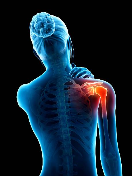
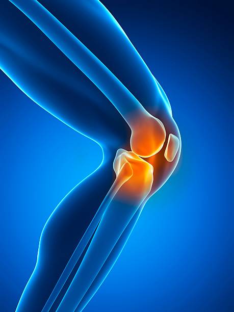
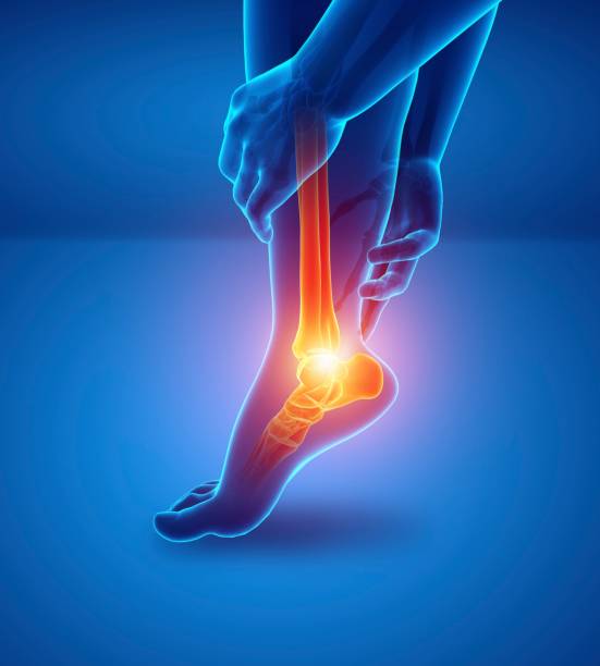
- Link to knee pain
- Patellar syndrome and associated pathologies
- Biomechanical analysis
- Clinical examination
- Orthopedic insoles
- Observe the mobility of the big toe in dorsiflexion
- The stretch test allows for a simple diagnosis
- Poor foot roll can lead to knee problems
- Correcting supports limits postural compensations
Information
Video type:
Anatomy:
Surgery:
Functional hallux limitus: understanding the starting point
Functional hallux limitus (FL) corresponds to a restriction of dorsiflexion of the big toe under load, not explained by structural osteoarthritis. This blockage disrupts the windlass mechanism and delays the transition from pronation to supination at the propulsive moment. This results in a lateralized roll, an external taligrade attack and chain compensations.
This dysfunction is not a local detail: it affects running economy and walking stability. Identifying HLF sheds light on the origin of pain sometimes attributed to the knee alone, when the initial problem is located in the forefoot.
Simple clinical diagnosis: the stretch test and walking
The stretch test consists of stabilizing the base of the first metatarsal, with the ankle in dorsiflexion, then pushing the hallux into extension. Failure to achieve dorsiflexion indicates a positive test. The examination is enhanced by clinical clues: absence of keratosis under the first metatarsal head, hyperkeratosis under the P2 pulp, and compensatory hyperextension of the interphalangeal joint.
Gait analysis confirms the phase shift: external heel strike, lateral support trajectory, then late medial tilt. These markers demonstrate excessive stress on the lateral structures and proximal desynchronization.
Functional hallux limitus is always associated with subtalar blockage.
Biomechanical chain: from foot to knee
The subtalar blockage prolongs pronation beyond the propulsive phase. The induced internal rotation causes a "sweeping effect" visible on the mat, with a late recentering of the axis. At the knee, the increase in varus stresses at the attack and then the medial collapse at the support reflect this shift.
This pattern explains some of the medial patellofemoral pain, goose foot tendinopathy, and iliotibial band pain. Walking becomes energy-intensive, and the muscles work in catch-up rather than in anticipation.
Injury mechanisms and sport: when timing goes wrong
The abrupt transition from supination to exaggerated pronation during the stance phase creates rotational shear that is unfavorable for the knee. This "highlight" corresponds to the window of vulnerability described in certain knee sprains, particularly in football or handball. The delayed contraction of the joint protectors increases the risk of injury, particularly to the anterior cruciate ligament.
Repeated lateral impacts increase stress on the internal compartment and the Achilles tendon. Recognizing the pattern helps guide prevention and rehabilitation specific to the sporting movement.
This sudden shift from pronation to supination can lead to knee injuries.
Patellar syndrome: from pain to mechanism
In HLF, quadriceps contraction occurs with a slight lag at the critical moment of step onset. The patella "floats" and then suddenly recenters, generating stress peaks and painful cartilaginous edema. The increased varisting moment and external rotation at onset explain the frequent association with band pain.
The examination often reveals amyotrophy of the vastus medialis, tension in the pes brevis and tenderness along the semitendinosus. Linking these signs to the HLF avoids treatments focused on the knee without correcting the distal cause.
Functional support: correct support, restore rhythm
The strategy combines education, weight-bearing correction, and neuromuscular training. Insoles can refocus the load on the first ray, while targeted exercises target hallux mobility, subtalar synchronization, and pelvic-femoral control. The goal is to restore the pronation-supination transition at the right time.
In this context, instrumental gait analysis and symptom monitoring guide adjustment. Correcting the starting point early limits the cascade of compensations and promotes a return to sporting activity without lasting pain.
Pathologies treated at the center
Hallux Limitus
Functional
Your pain has a cause.The balance sheet allows us to understand it.
- Gait analysis
- Posture Assessment
- Guidance on the right treatment
- Study of plantar supports and supports
- Detection of compensations
- Pain–movement correlation
The functional assessment allows us to understand how a joint or postural imbalance can trigger or perpetuate pain. Very often, imaging is normal, but movement is disturbed. By analyzing gait, weight-bearing patterns, or posture, we identify the weak links in the chain and guide targeted treatment adapted to the patient's actual mechanics.