Endoscopic tenolization for functional hallux limitus
In this video, Dr. Jacques Vallotton demonstrates a minimally invasive surgical procedure to treat functional hallux limitus. Using an endoscopic tenosynovitis device targeting the flexor hallucis longus tendon, the goal is to restore optimal mobility to the subtalar joint. This technique is particularly suitable for athletes with painful locking of the big toe.
Doctors
Topics
Treatments
Advice
- Dr Jacques Vallotton
- Functional hallux limitus
- Hyperkeratosis
- Endoscopic tenolization
- Subtalar arthroscopy
- Tendon release
- Endoscopic tenolization
- functional block of the hallux
- tenolizes to release the tendon
- subtalar joint
- common pathology among dancers
- minimally invasive treatment
Information
Video type:
Anatomy:
Surgery:
Thematic:
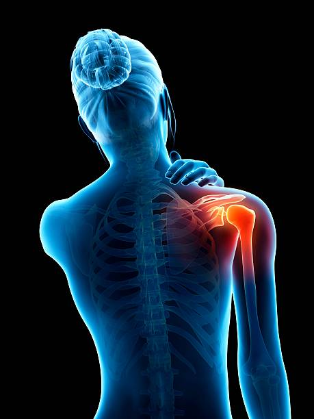
Functional hallux limitus: understanding the blockage
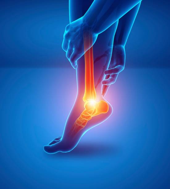
Functional hallux limitus manifests as painful blockage of extension of the big toe during walking, with suggestive cutaneous signs (pulpal hyperkeratosis, lateral transfer of weight bearing). The flexor hallucis longus (FHL) tendon inserts on the distal phalanx and slides in a tunnel posterior to the talus.
When this tendon becomes trapped at the retrotalar passage, the toe is no longer able to raise into dorsiflexion when the ankle itself is in dorsiflexion. This discomfort is common among dancers and skaters, who are subjected to repeated stress.
Endoscopic exploration of the hindfoot
The procedure begins with two mini-incisions, the introduction of the arthroscope and the creation of a working space on the posterior aspect of the subtalar joint. Landmarks include the lateral malleolus, the Achilles tendon and the posterior talocalcaneal ligament, which is sectioned without major impact on stability.
The examination allows the FHL to be identified, its sliding to be assessed and the retrotalar tendinous pulley to be located which bridges the posterior tubercles of the talus.
The long flexor tendon gets stuck at the tunnel and is unable to slide.
Tenolise: release the retrotalar pulley
Tenolization involves severing the fibers of the retrotalar pulley responsible for the conflict. The use of a hot dissection instrument followed by arthroscopic scissors allows for a complete and controlled release, while protecting the neighboring vascular-nervous bundle.
Once the pulley is open, the tendon is visualized along its entire length and tested in mobilization to verify the return of free sliding.
Mobility restoration and success criteria
Immediate success is judged by the recovery of dorsiflexion of the toe when the ankle is placed in dorsiflexion, indicating a tendon now freed in its tunnel. The subtalar joint regains full play at the end of the movement.
The minimally invasive technique aims for a rapid return to function with pain control and limited healing.
This tenolization will allow the joint to function again and no longer be fixed.
Indications, sports patients and prevention of recurrences
Endoscopic tenosynovitis is intended for patients with documented functional blockage that is resistant to conservative measures. In athletes, muscle hypertrophy and extreme stress promote impingement; management includes a gradual recovery program.
Proprioceptive work and load management limit recurrences, in addition to podiatric monitoring if necessary.
Care and rehabilitation pathway
Post-intervention rehabilitation targets gait pattern, rearfoot flexibility, and flexor/evertor strength. The goal is to restore efficient propulsion without proximal knee-hip compensation.
A clinical check verifies the persistence of free sliding of the FHL and adapts the progression towards sports supports.
Pathologies treated at the center
Hallux Limitus
Functional
Your pain has a cause.The balance sheet allows us to understand it.
- Gait analysis
- Posture Assessment
- Guidance on the right treatment
- Study of plantar supports and supports
- Detection of compensations
- Pain–movement correlation
The functional assessment allows us to understand how a joint or postural imbalance can trigger or perpetuate pain. Very often, imaging is normal, but movement is disturbed. By analyzing gait, weight-bearing patterns, or posture, we identify the weak links in the chain and guide targeted treatment adapted to the patient's actual mechanics.